Research Centers and Core Facilities

The Center for Molecular & Cellular Signal Transduction in the Cardiovascular System

UNR Med received Phase II funding of $11 million, for a second five-year installation of the grant from the National Institute of General Medical Sciences (NIGMS) of the National Institutes of Health (NIH) for a Center of Biomedical Research Excellence (COBRE) to study molecular and cellular signals regulating the cardiovascular system. This grant established Nevada's first cardiovascular-focused basic science center, supporting groundbreaking research in the field.


Translational Research Center

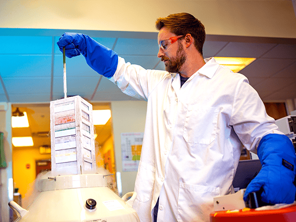
The Translational Research Center (TRC) is an on-campus, 5,470-square foot research space located in the Center for Molecular Medicine. The TRC offers private outpatient rooms for research clinic visits, two blood draw stations, sample processing and storage and much more. This modern research center has a variety of resources and on-site staff to support researchers and students who are making important strides in health care. The TRC is equipped with 11 clinical examination rooms, each of which include an exam table, otoscope, ophthalmoscope, vital signs equipment and sink. Researchers also have access to the following equipment:

  • Basic specimen processing area and a refrigerated centrifuge
  • Ambient, cold and frozen secure storage in two 2-8°C refrigerators, a -20°C freezer, and a -80°C freezer
  • Ice and dry ice in contiguous lab space
  • Automated vital signs monitor, AED, limited anthropometric equipment, secure document disposal, printer/fax/copier/scanner, analog line for data transmission

Research Core Facilities

Research at the University of Nevada, Reno School of Medicine (UNR Med) is supported by institutional core facilities that offer advanced technologies, state-of-the-art equipment, valuable resources and faculty expertise to advance the development of groundbreaking research at UNR Med. These resources are available to faculty and researchers across the University and to external collaborators.

Cell Analysis

The Cell Analysis Core Facility (CACF) is a shared resources facility that offers analytical fluorescence-based flow cytometry and cell sorting. Featuring a full staff and laboratory facility, CACF’s mission is to facilitate scientific progress at UNR Med by providing researchers with state-of-the-art resources and support to advance their research. Technical equipment includes:

  • A BD FACS Aria II SORP Cell Sorter with 4 Lasers (355nm UV, 488nm Blue, 561nm Yellow-Green, and 633nm Red) 16 Colors simultaneously, four nozzle sizes, inside a Biosafety cabinet for handling BSL2+ samples, Automated Cell Disposition Unit available for 96 or 384 well plate sorting
  • A Cytek Aurora Full Spectrum Cytometer with 5 lasers (355nm UV, 405 nm Violet, 488nm Blue, 561nm Yellow-Green, 633nm Red), Capable of handling 40 tubes automatically as well as 96 well plates or manual sample options
  • A Nanocellect WOLF Microfluidics Sorter with a single 488 Blue laser, optimal for cell culture post sorting, Capable of sorting into 96 well plates
  • A Beckman Coulter Cytoflex Cytometer with 3 lasers (405nm Violet, 488nm Blue, and 633nm Red), Ability to sample from Single tubes or 96 well plates
  • A Nanostring CosMx Spatial Molecular Imager
  • Formlabs Form 3B Resin 3d Printer
  • Prusa Filament 3d Printer

Biosafety Level 3 Lab

The Biosafety Level 3 Lab (BSL-3) is designed for researchers studying infectious agents. This lab is only available to University faculty who have had their protocols reviewed by Institutional Biosafety Committee.

Transgenic Animal Genotyping and Phenotyping

The Transgenic Animal Genotyping and Phenotyping Core (Core B) collaborates with investigators to provide services, consultation, training and technical support for evaluating transgenic animal genotyping and cardiovascular phenotyping research. This facility conducts cardiovascular research to facilitate the development of novel drugs for the treatment of diseases. The goal of Core B is to offer major cardiovascular phenotyping techniques including: continuous blood pressure, heart rate, ECG monitoring by radio telemetry system, in vivo calcium imaging and optogenetics in live free-moving mice, echocardiography and laser doppler blood flow recording. Available services and facilities include:

  • Breeding, genotyping and managing colonies of genetically modified mice to perform genotyping and data analysis of tail biopsy samples.
  • Monitor and record hemodynamic parameters including radio telemetry blood pressure, heart rate and ECG monitoring in up to 48 conscious, unrestrained mice.
  • Capture synchronized metabolic and behavioral information via the Sable Promethion Metabolic Cage System.
  • High-precision analysis and determination of fat mass, lean mass, free-water mass and total-body-water mass using the EchoMRI system.

High Spatial and Temporal Resolution Imaging

The High Spatial and Temporal Resolution Imaging Core (HSTRI) performs state-of-the-art high spatial and temporal resolution imaging methodologies for studying cellular structure, signaling pathways and function in health and disease. Recent microscopy advances have produced a new generation of instruments and molecular tools that enable biomedical researchers to investigate the relationships between structure and function in biological systems with unprecedented spatial and temporal resolution. These modern technologies allow visualization of the ultrastructure of cells and how it impacts localized signaling pathways. HSTRI trains, educates and advises investigators in the use of these new technologies to maximize the Core's impact on cardiovascular research. Available services and facilities include:

  • Supporting the following instruments: Leica Stellaris X8 confocal microscope, Leica Stellaris 5 confocal microscope, Olympus-Based Spinning Disc and TIRF confocal microscope, 4-Line Olympus Total Internal Reflection Fluorescence (TIRFM) and FRET Microscope System Combined with a Patch Clamp Workstation, Leica DMi8/SR GSD-3D Super-Resolution Microscope and 3i Lattice LightSheet Microscope.
  • Managing Imaris Software for advanced image analysis.
  • Sample preparation equipment for histology, immunohistochemistry and immunocytochemistry.
  • Development and troubleshooting of methods and standard imaging procedures, including assistance with advanced image analysis techniques.
  • Optimization of histology sample preparation for imaging such as sectioning and staining for fixed and live cell imaging.
  • Education of core users with novel microscopy and imaging techniques surfacing on the horizon.
  • Technical support and training of core users

University of Nevada, Reno - Biomedical Research Cores

Nevada Genomics Center

The Nevada Genomics Center is a state-of-the-art facility that is revolutionizing the study of genomes through advanced biomedical research. Located on the University of Nevada, Reno campus, our fully-staffed facility is equipped with the latest technologies and instruments to serve both academic and industry researchers. A team of experts is available to support investigators at every stage of their research in the function and structure of genomes. Our services support a range of options and sequencing yield, from pilot projects to intensive, data-rich projects.

Nevada Proteomics Center

The Mick Hitchcock, Ph.D., Nevada Proteomics Center offers high quality mass spectral proteomic analysis using the latest technologies and techniques. We aim to make our services accessible to both public and private entities including research faculty, students and health care professionals. Our team provides insight into the physiological state of cells to expand research on immune response and underlying health conditions. Our goal is to assist investigators with qualitative and quantitative proteomic analysis to gain a greater understanding of physiological pathways, molecular interactions and regulatory signaling.